Damaged Cartilage and Bone in Your Ankle
What is osteochondritis dissecans of the Talar Dome?
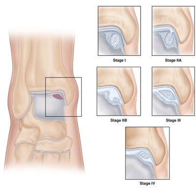
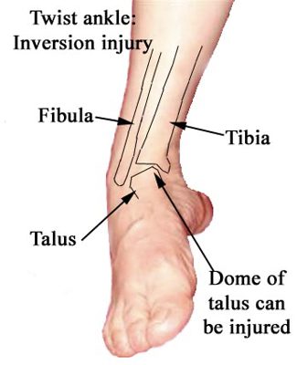
The talus is the lower bone in your ankle joint that supports the tibia and fibula. The talar dome is the top articular surface of the talus. Osteochondritis dissecans occurs when a small piece of bone begins to separate from its surrounding region due to lack of blood supply. This piece of bone cracks and loosens. The most common joints affected are the knee, ankle, and elbow. In advanced cases, the bone can completely detach and become a loose body in the joint.
How does it occur?
This is a condition that develops in joints most often in children and adolescents. Talar dome lesions usually occur with twisting injuries along with a sprained ankle.
What are the symptoms?
The most common symptoms are pain and swelling of the joint. In severe cases you may see joint catching and locking. You may be unable to bear weight on your ankle initially.
How is it diagnosed?
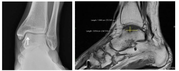
A complete history and physical exam will be done. Routine X-ray (left) of the affected joint will be done. This will evaluate the size and location. Then an MRI (right) will be performed to better evaluate the extent of cartilage affected and other injuries to the ligaments.
How is it treated?
In children with OCD, they can be treated with rest and physical therapy. They may heal on their own. If symptoms worsen, then nonsurgical means of treatment would be use of crutches with casting and /or splinting the affected joint.
Surgical treatment may be needed if symptoms don’t improve with time, if a lesion becomes detached, or if the lesion is very large.
Surgically you can drill holes to create pathways for new blood, hold the lesion in place with pins and screws, or by replacing the damaged area with a new piece of bone. This can be done arthroscopically or as an open procedure depending on the extent and location of the damage.
What is the recovery time?
Recovery depends on the severity of the injury. If surgery is required, it can be done as a same day surgery. Physical therapy with range of motion exercises will be helpful at this time. You will be in a non-weight bearing status using crutches or a walker for 6-8 weeks while healing occurs.
We are here to help. If you believe you are suffering from one of these conditions, we would love to deliver a diagnosis, get you treated and get you moving again.