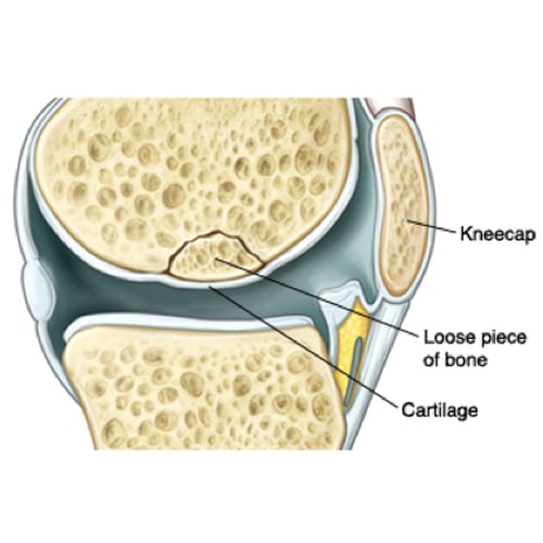
What is an Osteochondral Defect (OCD)
Osteochondral Defect is damage to the articular cartilage and the underlying bone that sometimes results in a piece of cartilage and bone coming loose into the joint space.
How does an Osteochondral Defect occur?
Disruption of the blood supply to these cartilaginous surfaces from vascular disease or trauma can weaken the areas. Repetitive trauma or stresses can also cause these surfaces to become damaged over time. The most common surface affected by an osteochondral defect is the end of the femur at the knee joint, but other commonly affected joints are the elbow and ankle.
What are the symptoms of an Osteochondral Defect?
Chronic pain and swelling of the affected joint are common after exertion. Eventually, locking or catching of the joint may occur.
How is an Osteochondral Defect diagnosed?
A history and physical exam will be performed. X-rays will help in locating the lesion and determining the size of the affected area. An MRI will also be taken to observe the soft tissue of the affected joint such as the articulating cartilage as well as the size of the lesion to assist in surgical planning. An MRI is essential to the diagnosis and management of treatment.
How is an Osteochondral Defect treated?
Rest and activity adjustments will be recommended initially on diagnosis to help with the pain and inflammation associated with an osteochondral defect. If the lesion is large or detached from the rest of the bone, surgery will be recommended to minimize complications and suffering of the affected joint. The defect can be repaired with transplanted tissue from a non-load bearing surface to the damaged area (called an osteochondral autologous transplantation (OATS) procedure). With a larger defect, an allograft from a donor cadaver must be used.
What is the recovery time after an Osteochondral Defect repair?
After an osteochondral defect repair, you can go home the same day as your surgery. Patients are shown simple range of motion exercises they can do at home, so there is no need to go to a rehab facility for the first 2 weeks, but once the graft has time to start healing, physical therapy may be recommended to help in the recovery process. Because the graft needs time to heal, patients will be on crutches for 6-10 weeks, but will gradually progress to normal activity after that with no high impact sports for 6 months.
We are here to help. If you believe you are suffering from an osteochondral defect, we would love to deliver a diagnosis, get you treated and get you moving again.